Metastatic liver cancer
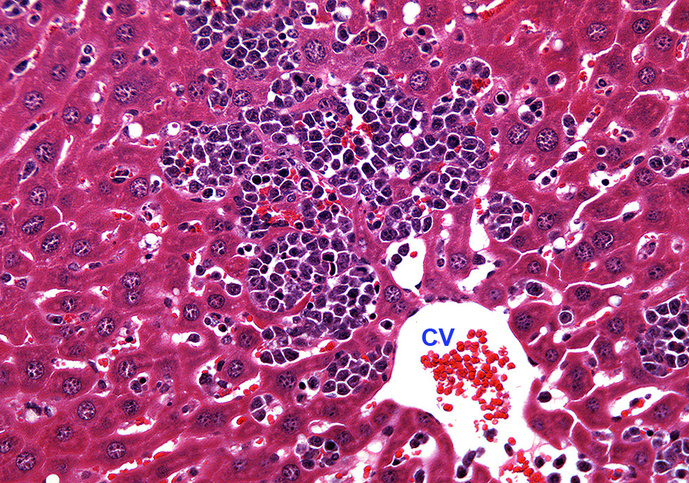 Histological image shows part of a mouse liver lobule with large cluster of abnormal mononuclear cells near the central vein (CV).
The liver plates (hepatocyte cords) among the tumor cells are mostly destroyed. Regular paraffin section with HE stain.
Histological image shows part of a mouse liver lobule with large cluster of abnormal mononuclear cells near the central vein (CV).
The liver plates (hepatocyte cords) among the tumor cells are mostly destroyed. Regular paraffin section with HE stain.