Skeletal muscle as seen with transmission electron microscope (TEM)
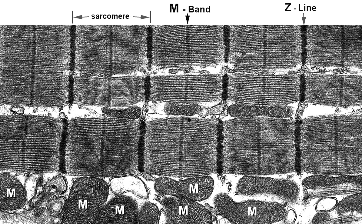
The skeletal muscle fibers consist of well organized functional unit – sarcomeres and other critical supporting structures such as the sarcoplasmic reticulum,
T-tubules, and mitochondria. This micrograph shows the typical structure of the sarcomere with the Z-Line and M-Band.
Each sarcomere unit is approximately 2μ in length and consists of paralelle myosin and actin filaments. Numerous mitochondria (M) are shown at
the bottom of the figure. Mouse skeletal muscle fixed with glutaraldehyde, embedded in Epoxy resin. Sections cut with diamond knife, stained with uranium and lead,
and examined with FEI Tecnai-T12 microscope.